Recovering from a total knee replacement (TKR) is a significant achievement, yet many patients find that regain-
ing a natural, symmetrical gait remains a challenge. Uneven walking patterns can impact mobility, confidence,
and overall quality of life. This case study explores how EMG can be a powerful tool to guide physiotherapy
interventions and significantly improve gait symmetry in a patient following TKR.
Patient Case: Addressing Gait Imbalance and Achieving Patient
Goals
Our patient, a 68-year-old female, underwent a left TKR due to severe osteoarthritis. Pre-operatively, each step
was a painful negotiation, a visible limp betraying the discomfort. Post-surgery, while the sharp pain subsided,
a nagging sense of imbalance persisted, making even short walks a challenge. She presented with decreased
weight-bearing on the operated limb, reduced step length, and a reliance on assistive devices. Her primary goals
were to walk without a cane, navigate stairs with ease, and return to her active lifestyle, which included gardening
and walking her dog. The initial physiotherapy assessment revealed weakness in the quadriceps, hamstrings, and
gluteal muscles on the operated side, contributing to the observed gait deviations.
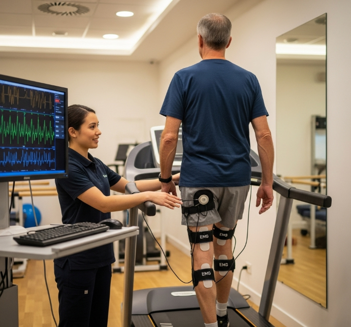
Unlocking Gait Imbalances: EMG Gait Analysis for TKR Recovery
EMG uses sensors to measure how well your muscles are working, helping us pinpoint exactly where the imbalances
lie. To gain a deeper understanding of the underlying muscle activation patterns contributing to the patient's gait asymmetry,
we employed EMG gait analysis using PHYSIOEMG. Surface electrodes were placed on key muscle groups, including the
quadriceps (vastus medialis and lateralis), hamstrings (biceps femoris and semitendinosus), gastrocnemius, and tibialis
anterior, bilaterally. The patient was then asked to walk at a comfortable pace on a treadmill while the EMG system
recorded muscle activity during different phases of the gait cycle.
The initial EMG data revealed several key findings. On the operated limb, the quadriceps muscles exhibited
delayed activation and reduced amplitude during the loading response phase, indicating weakness and impaired
shock absorption. The hamstrings showed compensatory overactivity during the mid-stance phase, likely to
stabilize the knee joint. Furthermore, the gluteus medius muscle, responsible for hip stability, displayed reduced
activation, contributing to a Trendelenburg gait pattern. These findings provided objective evidence of the muscle
imbalances contributing to the patient's asymmetrical gait.
Personalized Physiotherapy: Targeted Approach to Improving
Gait Symmetry
Based on the EMG findings, a personalized physiotherapy program was designed to address the specific muscle
imbalances identified. The program incorporated a multi-faceted approach, including:
• Strengthening Exercises: We prescribed targeted exercises to strengthen the weak quadriceps, hamstrings,
and gluteal muscles on the operated limb. These included isometric quadriceps sets, hamstring curls, gluteal
bridges, and hip abduction exercises using resistance bands. Progressive overload was applied to gradually
increase the intensity and challenge the muscles.
• Gait Retraining: Specific gait retraining exercises were implemented to improve weight-bearing, step length,
and cadence. These included mirror exercises, treadmill walking with visual feedback, and overground walking
with verbal cues to promote a more symmetrical gait pattern. Task-specific training was also incorporated to
enhance functional performance.
• EMG Biofeedback: We used real-time EMG biofeedback to enhance the patient's awareness of muscle
activation patterns during gait. Electrodes were placed on the quadriceps muscles, and the patient received
visual and auditory feedback on the level of muscle activity. This allowed her to consciously increase quadriceps
activation during the loading response phase, promoting improved knee stability and shock absorption.
• Neuromuscular Electrical Stimulation (NMES): NMES was applied to the quadriceps muscles to further
enhance muscle strength and activation. This involved delivering electrical impulses to stimulate muscle con-
traction, particularly during functional activities such as sit-to-stand and stair climbing. The application of NMES
is within the scope of physiotherapy practice for muscle strengthening and rehabilitation.
Outcomes: Quantifiable Gait Symmetry Improvements with EMG
Guidance
After eight weeks of consistent physiotherapy intervention, the patient demonstrated significant improvements
in gait symmetry and functional mobility. A follow-up EMG gait analysis revealed notable changes in muscle
activation patterns. The quadriceps muscles on the operated limb exhibited increased activation amplitude and
improved timing during the loading response phase. The compensatory overactivity of the hamstrings decreased,
indicating improved knee stability. The gluteus medius muscle showed enhanced activation, contributing to
improved hip stability and reduced Trendelenburg gait.
Clinically, the patient's walking speed increased, and her step length became more symmetrical. She was able to
wean off her cane and navigate stairs with greater ease and confidence. Her self-reported pain levels decreased,
and she reported a significant improvement in her overall quality of life. As our patient put it, "I can finally walk my
dog without worrying about falling. It's like I've got my life back!" Specific outcome measures, such as the Gait
Deviation Index and spatiotemporal parameters, were used to quantify these improvements. These objective and
subjective improvements highlight the effectiveness of EMG-guided physiotherapy in optimizing gait symmetry
after TKR. Furthermore, the normalization of the EMG gait cycle is important for accurate analysis.
Conclusion
This EMG case study underscores the potential of integrating advanced technologies like EMG into physiotherapy
practice to optimize rehabilitation outcomes after knee replacement. By providing objective data on muscle
function and guiding targeted interventions, we can empower patients to achieve greater gait symmetry, improved
mobility, and a higher quality of life.