Spinal stenosis can significantly impact your daily life, making even simple activities like walking or standing for
extended periods difficult or painful. The narrowing of the spinal canal puts pressure on the spinal cord and the
nerves that branch out from it, leading to a range of challenges. As physiotherapists, we understand the worries
and frustrations that come with this diagnosis. This blog post aims to illuminate the role of electromyography
(EMG) in understanding spinal stenosis. We'll explore how EMG helps assess muscle and nerve function, and
how we, as physiotherapists, use this information to create effective treatment plans tailored to your specific
needs.
Understanding Spinal Stenosis and the Physiotherapy Approach
Spinal stenosis involves the narrowing of the spinal canal, which can compress the spinal cord and the nerves that
extend from it. This compression can lead to pain, numbness, muscle weakness, and mobility issues. The location
of the stenosis (cervical or lumbar) influences the specific symptoms experienced. Our focus as physiotherapists
is to help you manage these symptoms, improve your function, and prevent the condition from worsening.
We achieve this through targeted exercises, manual therapy techniques like spinal mobilization and soft tissue
release, and comprehensive patient education. Patient education is crucial, including advice on posture, body
mechanics, and activity modification.
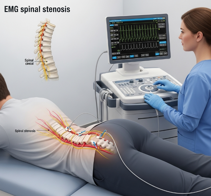
EMG's Role in Diagnosing Spinal Stenosis
In the context of spinal stenosis, EMG helps us determine if nerve compression is causing your muscle weakness,
pain, or other neurological symptoms. By measuring the electrical activity within the affected muscles, we can
gain insights into the severity and location of the nerve compression. The insights gained from EMG help
physiotherapists understand the impact of nerve compression on muscle function. The findings from an EMG can
reveal whether the nerves are conducting signals properly and whether the muscles are responding appropriately.
Specific electrical activity patterns, different from those in healthy muscles, can indicate nerve compression.
This information is crucial for differentiating spinal stenosis from other conditions that may present with similar
symptoms.
How Physiotherapists Use EMG Findings to Guide Treatment
The information obtained from EMG helps us tailor your physiotherapy treatment plan specifically for your spinal
stenosis. The findings help us pinpoint exactly which muscles and nerves are affected, so we can target our
treatments for maximum benefit. For example, if EMG reveals weakness in certain leg muscles due to nerve
compression, the physiotherapy program may include specific exercises to strengthen those muscles and improve
their function. Exercises might target specific muscle groups, with goals like improved strength and function.
Combining results from the medical history, physical examination, imaging tests, and EMG results, healthcare
professionals can accurately diagnose cervical radiculopathy and formulate a tailored treatment plan for each
patient. EMG findings can also inform the use of other physiotherapy modalities, such as electrical stimulation or
ultrasound. The treatment plan is adjusted based on the patient's progress and repeat EMG findings.
The Physiotherapy Assessment and EMG
The physiotherapy assessment is a comprehensive process that involves several key components. First, we
gather information about your symptoms, including their nature, duration, and any related incidents. We ask
detailed questions to understand your experience. Next, we conduct a thorough physical examination, assessing
your range of motion, strength, and neurological function. We perform specific tests to identify areas of pain or
weakness. We may also utilize imaging tests, such as X-rays, MRI, or CT scans, to visualize the spine and identify
any structural abnormalities. Finally, EMG is used to measure nerve and muscle function, helping us to determine
the extent of nerve damage or dysfunction. Patient-reported outcome measures also help assess the impact of
spinal stenosis on daily life.
Conservative Management of Spinal Stenosis
Managing spinal stenosis often involves a combination of physiotherapy, ways to manage your pain, and changes
you can make to your daily life. Physiotherapy plays a central role in improving function, reducing pain, and
preventing further progression of the condition. Clinical examinations also help indicate positive straight leg raise
tests, diminished Achilles reflex, and sensory deficits along the sciatic nerve distribution. Treatment approaches
may include exercises to strengthen core and back muscles, stretching to improve flexibility, and manual therapy
to relieve nerve compression. Pain management strategies may include medication, injections, or alternative
therapies. Lifestyle modifications, such as weight loss and ergonomic adjustments, can also help. Persistent pain
despite treatment with analgesics, non-steroidal anti-inflammatory drugs (NSAIDs), or muscle relaxants can be
a sign of failure of conservative treatments. Bracing or orthotics may also provide support.
Conclusion
EMG is a valuable tool in the diagnosis of spinal stenosis, providing crucial information about nerve and muscle
function. Understanding how EMG helps diagnose spinal stenosis empowers you to take an active role in your
care. The key to successful outcomes is the collaborative approach we take: your doctor interprets the EMG,
and we use that information to guide your rehabilitation. Failure of Conservative Treatments is a sign of the need
for further intervention. The long-term benefits of physiotherapy in maintaining function and preventing symptom
recurrence are significant.